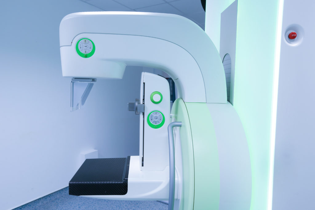
3-D Mammograms: The Next Big Development In the Fight Against Breast Cancer

Statistics show that annual mammograms are one of the best ways to detect breast cancer in its earliest stages, when it is most treatable. Now, a breakthrough diagnostic tool called 3-D mammograms is providing improved imaging and accuracy. Los Angels-area OB/GYN Dr. David Ghozland is among the first to offer this revolutionary new scan.
“A recent large study published in the Journal of American Medical Association indicates that 3-D mammograms may be better at finding cancer than regular scans,” says Dr. Ghozland.
Standard mammograms are able to show changes in the breast up to two years before a patient or physician can feel them, which is why doctors urge women to have annual screenings. Mammograms can also prevent the need for extensive treatment for advanced cancers and improve chances of breast conservation. The new 3-D mammogram, when used in conjunction with a regular mammogram, detected one additional cancer per 1,000 scans, compared with conventional digital mammograms. The study showed there were 15 percent fewer false alarms—questionable findings that lead to additional and ultimately unnecessary testing.
The study involved nearly half a million breast scans, with more than one-third of them using relatively new 3-D imaging. Among the advantages:
- 3-D scans may find more invasive forms of cancer at an earlier stage.
- The scans themselves take only a few seconds longer than traditional imaging.
- 3-D scans take several images of different layers of each breast, and may detect tumors hidden beneath breast tissue.
- 3D mammography may be particularly effective for women with dense breast tissue or those at high risk for developing breast cancer.
To understand the difference in the new technology, it’s important to note that traditional mammography produces two images of each breast, a side-to-side view and a top-to-bottom view. With a 3D mammography, Los Angeles physicians like Dr. Ghozland can obtain numerous X-ray images of the breasts from multiple angles; the resulting in a digital 3-dimensional rendering of internal breast tissue. With this type of detailed imagery, a radiologist can view the breast in 1-millimeter segments rather than observing it in full from the top and from the side.
While long term studies are still being done, current research suggests that the advantages of 3-D mammograms are numerous. The bottom line, says Dr. Ghozland, is that more detailed images will lead to fewer false-positive and false-negative readings, and ultimately, a more accurate diagnosis.
